w

Adenomatoid Odontogenic Tumor (AOT): Definition, Clinical features, Diffrential diagnosis

Definition 

The adenomatoid odontogenic tumor is a relatively uncommon,well-circumscribed,odontogentc neoplasm charactezed by the formation of multiple ‘ducts-like’ structures by the neoplastic epithelial cells.

The name ‘adenomatoid’ has been given to the neoplasm because histologically numerous duct-like are often intersersed thorough out the lesion give glandular or matoid apprarance to it.

Origin

The tumor probably arises from the reduced enamel epithelium,during the presecretory phase of enamel organ development.Some investigators believe that the neoplasm develops from a pre-existing dentigerous cyst.

Clinical Features

Age-The tumor usually occurs in the younger age (e.g.second and third decade of life) Rarely it can occur in the older age.

Sex-Females are more  commonly affected in comparison to the males.

Site-The lesion most typically occurs in the maxillay amterior region (upper laterag incisor-canine area) Rarely it involves the mandeble in the angle -ramus area in about 70% cases the neoplasms occur in association with an unrupted tooth , Some lessions develop extra orally in relation to the gingiva.

Clinical Presemtation
AOT Enucleation
AOT Enucleation Surgery
Image via: www.dental.washington.edu

  • The tumor usually presents a slow enlarging , small ,bony hard swelling in the maxillary anterior region.
  • Sometime it can occur in the premolar region k.
  • The lession often causes elevation of the upper lip on the involved side ,which often resuts in a change in the facial profile.
  • Dislacement of the regional teeth , mild pain and expansion of the cortical bone are usually present.
  • If the lesion is very large it may cause severe expansion of the which may sometime elicit fluctuation.
  • In many cases, the lesion is asymptomatic in nature and it is often associated with an uperupted tooth (mostly the upper canine)
  • Occasionally adenomatoid odontogenic tumor may occur extra-ossously in the anterior maxilly gingiva and it prodeces a solirary painless , asymptomatic nodular swelling. 
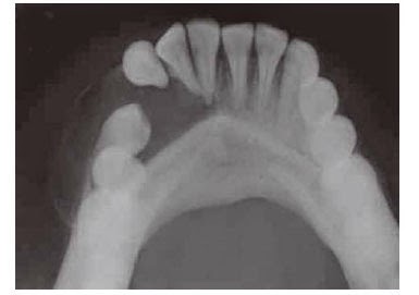
Radiological Features
AOT Radiologic feature
AOT of mandibular left canine region
image via: jaypeejournals.com
  • Radiographically adenomaroid odontogenic tumor presents a well-defined , unilocular,radiolucent area,which is often enclosing a tooth or tooth-like stucture.
  • Multiple small,radiopaque foci of varying radiodensity may be predent inside the lesion .
  • Expansion of the distrotion of the cortical plates and displacement of the roots of the adjoining teeth are sometime seen.
  • The border of the lesion is not well corticated and it consistently engulfs the impacted tooth including its root.This feature differentiates adenomataid adontogenic tumor from dentigerous cyts,since the later lession encloses only the crown portion of on impacted tooth.

Differential Diagnosis

  • Dentigerous cyts
  • Globulomaxillry cyts
  • Lareral periodontal cyts
  • Odontome
  • Unicystic ameloblastoma
  • Ossiffying or cementifying fibroma
  • Calcifying epithelial odontogenic tumor
  • Calcifyinh epithelial odontogenic cyts.


Histopathological Feature

  • Microscopically, adenomatoid odontogenic tumor reveals neoplastic odontogenic epithealial cells,proluferating in multiple "duck-like" patterns,within a thin but well-vascularized stroma.
  • The presence of these duck-like strucrures often give the lesion an adenomatoid or glandular apperance.
  • Each dick-like structure is bordered on the periphery by a single layer of tall columnar cell resembling ameloblasts.
  • Serial sectioning reveals that the lumens are blind ended ang they probably represent an abortive attempt at anamel oraga formation.
  • The lumen of the duck-like structures are filled with a homogenous eosinophilic coagulum.
  • Small foci of calcification are often seen , which are scattered throughout the lesion.
  • In some cases , the neoplastic cells are arranged in solid nests or rosettem and these cells simetime may fill up the entire lumen of few ducts.
  • Droplets of amorphous (PAS prositiv) eosino philic materials are frequently found in between the neoplastic calls.
  • In some adenomatoid odontogenic tumos tubular dentin or enamel matrix may also be found.
  • The neoplasm is almost always well-encap sulated and the connective tissue stroma may occasionally contain diffuse areas of hyaline materials.


Treatment 

By surgical enucleation . The associated tooth has to be removed and recurrence is rare.


Can Xylitol Chewing Gum Protect Your Teeth?

Xylitol
image via: Vitalsurge.com
You have always heard that eating sugar candy is bad for your teeth. but what if i say "chew some gum and protect your teeth"

Yes you are hearing right, i am suggesting you chewing gum ..but how does it help protect your teeth?..

I am suggesting you to have SUGAR FREE Xylitol containing chewing gums present in the market in various brands.


How does it help.?
As I have discussed earlier about the process of Caries/Decay, the organism responsible for decay of the tooth is S mutans.It promotes an acidic environment in our mouths which leaves enamel susceptible to damage, erosion and eventually cavities.

Luckily, nature has provided a cheap, natural and safe remedy for this problem: xylitol. xylitol is a sweet substance commonly found in birch trees and in the fibrous portions of many fruits and vegetables.I want to discuss its application as a cavity fighting agent.

A recent study out of Korea examined the effect on cavities caused by the regular chewing of xylitol sweetened gum.

Two groups of women were assigned to chew either regular or xylitol gum for a period of one year. At ten points during that year, saliva samples were taken from these women and analyzed to determine the amounts of S. mutans.

In the xylitol gum group, the levels of S. mutans decreased consistently as the study progressed.  The researchers also found that the S. mutans produced a lower amount of sticky substances in the xylitol chewers. This is relevant because the stickiness allows for acids to cause more damage to our teeth. The combined effects of  chewing the xylitol gum led to an oral environment that was less prone to cavity formation.

The results of this first study were encouraging. But I wanted to see if this might be a fluke. It seems that it is no

Facts & Figures-
In December of 2008, a review appeared in the Journal of the American Dental Association. In it, the researchers examined the findings of 19 studies relating to the use of xylitol and sorbitol gums in the prevention of tooth decay. Their analysis found that the xylitol gum studies showed the greatest cavity prevention.

Here’s a breakdown of a few different sugar alcohols and their overall preventive effect:
Xylitol Gum – 58.66%
Xylitol & Sorbitol Combination Gum – 52.82%
Sorbitol Gum– 20.01%
Sorbitol & Mannitol Combination Gum – 10.71%

As you can see, the xylitol component appears to be the most important factor in the promotion of oral health.

So how exactly does xylitol help protect teeth? It is believed that it works to starve harmful bacteria, like S. mutans. This leads to a less acidic environment that is less prone to decay and plaque formation.

Calcium added Xylitol gums-
Preventing cavities is a very positive thing. But is there a way to strengthen enamel, if it’s weak to begin with? Maybe so.
A few years ago, scientists in Japan published a study that tested a combination gum that included calcium lactate and xylitol. Their aim was to see if such a gum could actually make tooth enamel stronger.

Volunteers were asked to either a) chew no gum, b) chew xylitol gum or c) chew gum with xylitol and calcium lactate. The voluteers chewed 4 pieces of each gum for 2 weeks. After which, their enamel was measured using an X-ray.

The results showed that the xylitol-calcium gum was about 50% more effective in promoting remineralization than the xylitol-only gum. The authors concluded that, “chewing gum containing xylitol + calcium lactate could enhance remineralization of enamel surface”.
So i suggest having these xylitol chewing gums instead of sugar candies. Care for your teeth they are precious.


What is Sialolithiasis –Definition, Pathogenesis, Clinical features, Treatment, Histopathology, Sialography.

Sialolithiasis

Definition- sialolithiasis is a pathological condition, characterized by the presence of one or more calcified stones (sialliths) within the salivary gland itself or within its duct.

Pthogenesis

The exact mechanism of formation of sialolith is not known. It is generally believed that initially a small and soft nidus forms within the salivary glands or its ducts due to some known reasons.

The nidus is made up of mucin, protiene acteria and desquamated epithelial cells.

Once a small nidus frms, it allows concentric lamellar crystallizations to occur due to the precipitation of calcium salts.

The sialolith increases in size with time as layer after layer o salts become deposited , just like growth rings in a tree.

Small sialoliths can be expelled in the mouth with the salivary secretions but bigger sialoliths continue to expand until a duct is completely closed.

It is important to note that the formation of sialolith is more common in relation to the submandibular gland and it’s duct.

sialolithiasis

Clinical features

Age- Sialolithiasis usually occurs among the middle aged adults, however, some cases are reported in children.
Sex- There is slight male predominance.
Sites- 70% in submandibular salivary gland and its ducts. Parotid gland is next

Clinical Presentation-

  • In many cases sialoliths do not produce any symptoms and are detected only on routine radiographic examination.
  • Chief complain ; intermittent pain, recurrent sumbandibular swelling, discomfort during meals.
  • Pain is drawing or stinging in mild cases but stabing and sharp in severe cases.
  • Stone in the duct can be palpated by bimanual palpation with both the fingers.
  • During examination the flow and clearness of the saliva should be checked.
  • Persistent swelling of the duct due to chronic obstruction by the sialolith eventually leads to chronic sclerosing sialadenitis.
  • Multiple stone or bilateral cases of sialolithiasis can also be seen.
  • Formation of fistulas, sinus tract, ulcerations in the area may develop in chronic cases.
  • Necrosis of gland acini and lobular fibrosis may occur which results in complete loss of secretion from the gland.
  • Parotid stones often cause firm swelling over the ramus of the mandible, the swelling over the ramus of the mandible, the swelling also increases during meals.

How Sialolithiasis is diagnosed
  • Radilography of sialolithiasis
  • Submandibular sialolithiasis are easily detected by mandibular standard occlusal radiograohs which typically disclose the presence of calcification in the floor of the mouth.
  • Whn a sialolith is located in submandibular gland area then a lateral jaw radiograph will be helpful in location the exact position of the stone.
  • Panaromic radiographs usually detect the parotid gland stones.
  • If only branches of the submandibular gland duct are affected a posterior occlusal film submento vertex and some time a lateral jaw film may be required.

 Sialography of sialolithiasis
  • Sialogrohy refers to the method by which detection of salivary stones within the glad or its duct is done by giving a retrograde injection of a radiopaque dye within the  duct system and obtaining a radiograph thereafter in order to see the size and distribution of the sialolith.
  • Ultrasonograhy and CTscan
  • These are also excellent non invasive technique useful in detecting the sialoliths

Macroscopic appearance
  • On gross examination, sialoliths appear as round or oval rough or smooth solid masses, which vary considerably in their size.
  • These stones are heavily calcified and are often multi nodular although some stones are found in small aggregates.
  • The color of the stone is usually yellowish or yellowish white.


Why Are My Teeth Getting Stained ?

Question- I go to my dentist regularly and get my oral prophylaxis & cleaning done in every 6 months, My teeth become pearly white after cleaning and their are no stains but they start getting stained in no time ! I can see visible stains after 3 weeks.Why are my teeth getting stained even when I brush properly.

Although teeth staining have multiple causes but the above case is a simple case of poor oral hygiene practice. I'm not saying that the person is not will to keep his teeth clean but he is actually practicing wrong oral hygiene techniques like not knowing proper brushing technique, not flossing and brushing only once in the morning.

Cause of teeth getting stained after scaling and cleaning:
teeth getting stained
Stained between teeth Img: www.smileartistry.com.au

After professional cleaning surface of your teeth become absolutely clean but as you start eating a soft Dental pellicle start to form on your teeth.It is a protein film that forms on the surface enamel by selective binding of glycoproteins from saliva.

When not cleaned and flossed properly oral bacteria start to make colonies on this pellicle layer and hence they for a layer called as Plaque. This plaque can take the colors of foods and beverages your consume and it looks like a staining on your tooth.when you get your teeth scaled and cleaned at the dentist he removes this plaque. When people does not get this Plaque cleaned and it keep on accumulating it becomes Calculus which is a calcified structure.

Foods & Habits That Cause Your Teeth to Get Stain


Tea Or Coffee:
Tea Or coffee contains such kind of plant extracts that have very high tendency of sticking as stains on your teeth. No matter what type pf tooth paste you use, no matter what type of brushing you do, if you are consuming tea or coffee more then once a day every day in a year then your teeth will catch stain.Either get a professional cleaning or change your habit.

Smoking: Tobacco smoke contains nicotine and tar these two ingredients are the main culprit for the smoking stains. tar has a black staining and nicotine has a yellowish staining property. So a smoker will have a blackish and line at the junction of the gums and tooth and yellowish hue on the teeth. Leave the habit, Get your teeth cleaned.

Red Wine- Red wine is made up of grapes which have natural colors these natural colors will bind with the tooth surface and result in teeth staining, but this process is low and mild and occurs to them who consume red wine regularly.

Some Cold drinks and Energy drinks: Cold drinks and energy drinks have coloring agents in them. These agents can deposit on the plaque of the tooth and then get solidify.

Some berries and veggies: I really don't think that veggies should be blamed for the staining of teeth but clinically it have been seen that there are some berries and veggies which have colored juices in them which can stick to the plaque and impart

Solution to Get Rid of Repeated Teeth Staining

Avoid stain producing habits and food.
Brush twice a day with correct brushing technique.
Use floss once a day.
Use inter dental brush to clean gaps between teeth.
Get oral prophylaxis done every 6 months.
Keep oral hygiene good.

What is The Blue Light Dentist Use : Is It Harmful ?

So you have been to the dentist and you saw a wand like thing in dentists hand which he puts in patients mouth then it emits bright blue light with sound of beep !

No need to wonder what is that blue light emitting thing in dentists office.

 I'll Explain what is it, how it works and is there any chance of harm from it.

Blue light that dentists use is called as curing light.

How does blue curing light works ?

Blue dental light
Image Via : Wikimedia.org
It is a gun like machine which have electronic parts inside it. The source of current is the regular 220 volt AC current but a chip inside the machine converts it into much lower DC current and then it supplies it to the light emitting source.( Some guns are battery operated)

The light emitting source is a Quartz Halogen Or LED ( Light emitting diode) attached to the head of the gun. LED bulb have the capacity to produce high intensity of light with very effective use of electricity with least heat production as compared to other bulbs. hence a LED is used. (Don't miss judge it with lasers or any other thing) .

To make an angulated head a fiber optic cable is fitted at the head ahead of the LED bulb, this fiber optic cable can carry light to the head even when angulated at various angles.

Why is the dental curing light blue in color ?

It is mostly blue in color because the light is of longer wavelength which helps in deeper penetration of the light for better curing of the material. The wave length of the light may range from 370 to 510 nm. This light falls in blue spectrum of the VIBGYOR.

What is the blue curing light used for ?

This light is used for setting light activated filling materials.
In Technical Terms- There are dental restorative materials which come in semi solid or powder liquid form when mixed and packed into the cavity they get activated and start setting. Some material are photosensitive- That means setting reaction is initiated after light exposure. Blue curing light is used to cure this kind of dental materials.


Is the blue light harmful ?


No it's not harmful for the tissues inside your mouth or other skin or mucosal tissue, BUT it should never be directed to the eye or in other words you should not look directly into the light, because it have so much of intensity that it may harm retina of eye.

What is a orange colored shield ?

Due to the above cause there is a light barrier or orange color which dampens the light intensity if the operator is working and want to see the field of work.  For patients a orange colored glass will be given, if not given you should better keep your eyes closed.

Why Are My Teeth Getting Sensitive

Teeth getting sensitive means you are feeling sharp electric like pain on taking cold, sweet or sour food, this kind of pain is short in duration and it subside immediately after removal of stimuli.

If that is what you are experiencing then you are suffering from a clinical condition called as Dentin Hypersensitivity Or Sensitive Dentin.

Teeth crown is made up of outer Enamel layer (Don't have nerves), Dentin layer (have nerve ending in it) & Core of Pulp (nerves and blood vessels). Teeth root is made up of outer Cementum and then Dentin layer.

These enamel and cementum works as insulation to underling dentin from temperature and osmotic stimuli. So now you can figure it out that when ever enamel or cementum is damaged and dentin is expose people start to feel sensitivity while eating cold or sweet food.

Causes of Teeth getting sensitive:
Teeth Getting Sensitive
Picture showing Causes of Sensitive Teeth 


Gingival recession or receding gums: This is most common cause of teeth sensitivity gums cover the root which intern is covered by the cementum. when gums recession occurs then root get exposed and by the time when cementum is removed from brushing then tooth become sensitive. Heavy brushing and periodontal disease are two causes or gum recession.

Tooth wear: This is second most common cause: As you know that wear and tear is a natural process but when this process occurs at high speed and your enamel is lost at fast speed then your teeth does not get time to perform any kind of protective mechanisms. Hence tooth become bad before time and you can experience tooth sensitivity. Example of tooth wear- Attrition from teeth to teeth contact ( physiologic), Abrasion ( tooth brush trauma- Pathologic), Abfraction ( combination of both), Erosion ( chemical damage from the acid erosion of the enamel from consuming excessive cold drinks, Gastro esophagial reflux and many more)

Dental decay: Yes decay can also cause teeth sensitivity as decay causes destruction of enamel it exposes the dentin and when ever cold or sweet food comes in contact with that decayed tooth patient may feel sharp pain. which subsides on removal of the stimuli.


Treatments: Depend upon the grade and cause of sensitivity.

Removal of cause and treatment of underlying pathology followed by Desensitizing toothpaste, Desensitizing gel & Professional Dental treatment will completely cure your sensitive Teeth.

You can Check These Desensitizing Tooth Pastes If you want one.

Cauliflower Like Growth in Mouth !

Cauliflower like growth in the mouth is commonly due to papilloma. It's a benign neoplastic growth which is usually not fatal.

Papilloma definition-  Papilloma is a common benign neoplasm arising from epithelial tissue of oral cavity and is characterized by an exophytic growth of the stratified squamous epithelium.

These lesions  constitute about 2% of all oral neoplasms and it is believed by many investigators that they are caused by the human papilloma virus.

Clinical features

Age: mostly third, fourth and fifth decade of life
Sex: Both sexes are equally affected.
Site: Tongue lips, buccal mucosa, gingiva, hard and soft palate.

Signs and symptoms: Clinical presentation


  • ‘Cauliflower like’growth which is slow growing exophytic and small, it is usually painless unless traumatized. It has several small finger-like projections on the surface. 
    Cauliflower like growth
    Papilloma on roof of the mouth.
  • Size is from millimeter to few centimeter in diameter.
  • The base is either pedunculated or broad based (sessile) but always well-circumscribed lesion.
  • Sometimes it may appear as ovoid swelling with the corrugated surface.
  • The surface of the papilloma is white because of the highly keratinized epithelium.
  • A non-keratinized lesion appears pinkish or grayish in color and it is softer in consistency.
  • Rarely an endophytic papilloma can be seen which grows inwardly.
  • Papillomatosis is a condition in which multiple papillomas are developed inside the mouth.


Histological feature
Papilloma
Histological slide of papilloma


Proliferating stratified squamous epithelium in the form os multiple fingers like projections

Every single finger-like process has a vascular connective tissue core in cener which conatins few inflammatory cells.

Covered with squamous epithelium which shows hyperkeratosis. and acanthosis.

Although most of the papillary projections are long and finger like there can be small round and blunt projections.

Sometimes there may be a development of superficial ulcerations and secondary infection, because in the oral cavity these lesions are often subjected to trauma.

Differential diagnosis

  • Verruca Vulgaris
  • Focal dermal hyperplasia
  • Verruciform xanthoma
  • Verrucous carcinoma 
  • Fibro epithelial polyp.


Treatment

Surgical excision of the lesion including the base. Recurrence is rare.

What is Hairy Tongue: Pathogenesis Clinical Features and Treatment

Definition Hairy tongue is an unusual condition which occurs due to hypertrophy of the filliform papilla of the tongue along with loss of normal desquamation process This eventually leads to the formation of a pigmented thick matted layer on the tongue surface heavily coated with bacteria and fungi.

Pathogenesis: Why hairy tongue occur 
Hairy tongue
Hairy tongue

Normally the keratinized surface layers of the tongue papillae are continuously desquamated thourg
friction of the tongue with food, rough palate and upper anterior teeth. After desquamation tongue papillae are replaced by newer epithelial cells from below.

Lack of tongue movement due to local or systemic disease disturb the regular shedding of the keratin of the tongue papilla especially the filliform papilla.which lengthens and produces a hairy appearance on tongue surface.

Such hypertrophic papillae are often coated with microorganisms and become discolored by retaining pigments from foods, medicines and chromogenic bacteria etc.

Clinical Features of hairy tongue how does it look


  • Hairy tongue commonly affects the mid dorsum of the tongue.
  • Hypertrophy of the filliform papilla produces a thick matted layer on the dorsal surface hairy tongue in extreme cases may produce a thick lathery layer coating on the tongue surface and this condition is often known as early or encrusted tongue.
  • Hypertrophied filliform papilla often brushes sfot palate and produces gagging reflex.
  • Hairy tongue in many cases produces bad breath.
  • There can be irritation to the tongue due to accumulation of food debris and microorganisms.
  • Hairy tongue is considered as mirror of general health. Because it is associated with various systemic disease.


Treatment of hairy tongue.

  • Cleaning and scraping of tongue everyday
  • Application of topical keratolytic agents
  • Consumption of yogurt
  • The effected tongue papilla often rapidly returns to normal when long term antibiotics or oterh drugs are discontinued.


What Is Laser Filling For Treating Tooth Cavity

laser fillingDental cavities are becoming inevitable and dental restorative procedures are become more advanced day
by day. Laser filling is one of such advancement.The most commonly used lasers in dentistry are ND- Yag laser, Helium Neon laser, Ruby Laser.

LASER stands for Light Amplification by Stimulated Emission of Radiation.

In simple language a laser filling is a restorative procedure in which the decayed portion of the tooth is removed with the help of specially designed dental laser and then it is restored with the help of tooth colored cosmetic filling.

Details: 
The objective of any kind of dental filling is same:- Remove the decay and restore the tooth in biocompatible

and functional manner. Laser filling also follow these basic steps.

Step one: Take an X ray of tooth and see that cavity should be small and it should not be close to the pulp (nerve) of the tooth.

Step two: Remove the carious lesion using dental laser. A water jet is continuously flowing while the laser is removing the decayed tooth material from the cavity. This kind of removal does not produce vibration or heat while removing the cavity hence patient comfort is good and easily achieved.

Step Three: After removing the decayed portion of the tooth the cavity is disinfected by using chlorhexidine gluconate  solution. Cavity is irrigated and dried for the filling.
dental laser
Dental laser

Step Four: A biologically compatible restorative material like light cured GIC or Composite resin is used for filling and binding the prepared cavity. After placing and curing the restorative material normal anatomy and function of the tooth is restored.

Step Five: Occlusion is checked with the help of occlusal bite registration paper and high points are removed.

What are advantages of laser filling

No vibration because no use of drill or burs this makes it more comfortable and  easy for patient.
No Shot of anasthesia is required: Actually it depends upon the pain tolerance of the patients but in most cases no shot is required.
Fast: It really takes few minutes to complete the removal of caries and do the filling.

What are the drawbacks of laser filling

Can't be used in large cavities
Expensive
Can be performed by very few trained dentists ( Lack of availability)


What is a Dental Mirror Used For

Dental Mirror is a small round mirror placed on a handle with variable degree of angles to it. These mirrors can have different convergence like concave, flat or convex but mostly concave is used.

What is a dental mirror used for? 

There are three main uses of dental mirror by dentists.

1. Indirect vision- It is obvious to understand that there are lot's of areas in mouth where direct vision is not possible, Either dentist have to bend upside down or patient have to get fully supine and extend back which is very uncomfortable situation for both of them so mouth mirror will help. A dental student will learn dentistry by learning to work in indirect vision using mouth mirror or so called dental mirror, And they really deserve some respect.


Dental Mouth Mirror
Indirect Vision & Illumination with mouth mirror Img Via www.hu-friedy.com

2. Indirect Illumination- Same reason given above it is difficult to illuminate every deep and detailed areas of the mouth with dental chair light. So the dental mirror is used for illumination of the particular areas of mouth by reflected light from dental chairs light. Use of concave dental mirror really help in this kind of illumination.

3. Retraction- Retraction in dental surgery means retracting the surrounding tissue like tongue and cheek to expose the areas for proper vision and accessibility for instrumentation. Retraction of cheek and tongue is easy with help of dental mirror,  just place the mirror side of the mouth mirror toward the tissue and pull it away from the site of operation. As the mirror is smooth that is why patient don't feel irritation. A trained dentist will quickly switch between illumination, retraction and vision while doing a dental treatment. Again Respect For These Artists.

There are many ways for gripping and holding mouth mirrors like pen grasp, three finger grasp etc. but that is out of scope of this article, Various types of  mouth mirrors are present in the market today but No4 and No5 are used mostly with concave mirror on it.
So Now yo know What is a dental mirror used for Now share it with your friends using share buttons bellow
You can buy same kind of kit for cleaning your teeth from Amazon here.

What is a Dental Milling Machine

Dental Milling Machines are machines used to produce dental prosthesis like crowns and bridges and implant prosthesis by the process of computer added designing and computer added manufacturing process CAD CAM. As the name suggests dental prosthesis are made by milling a block of ceramic or zerconia or metal aloy into desired design.

Use of Dental milling machine have been introduced in 1980 but real use has started from 1990, in early days they have been cumbersome to use and too big to handle, this restricted its use to dental labs.
Dental milling machine
Dental Milling Machine

Components of Machine- Machine basically includes a Scanner, Computer and Milling Machine.

Scanner which scans the area of teeth and mouth on which prosthesis is going to be build. If scanner is

present at the chair side in a dental clinic then scanning can be done directly in mouth, if scanner is not present chair side then an impression is taken and model is prepared and then scanning is done on the dental model.

Computer and software do all the calculation and designing of the dental prosthesis. It takes the data from scanner and builds a 3 D picture of it then builds a prosthesis on that #D picture this data is then given to the milling machine so that it can start milling and start manufacturing the prosthesis.

Dental Milling Machine is a fitted with small heads which includes different kind of burs or milling points.
these are diamond coated heads of different shapes and designs made for different purposes of milling.


How is it different from older conventional dental crown and bridge prosthesis.

  • Dentist will first examine your mouth and diagnose the disease and problem.  
  • He will treat the defect by removing decay and treating infections by root canal treatment. 
  • In third step dentist will prepare the area for receiving prosthesis. 
  • He may take and impression for making model or he may directly scan the portion on which prosthesis is going o be made.
  • This dental model or scanned data is send to lab where it is feed to computer and then it makes a digital 3 D model of that scanning.
  • digital prosthesis is prepared on that 3D model and then this data is given to the milling machine. 
  • Milling machine prepares the exact replica of the 3D design.
  • This prosthesis is send back to dentist for checking and applying in patients mouth. 
A Video of complete process From Datron Dental Milling Machine


9 Reasons And Causes for Chipping of Tooth : Infographic

Chipped tooth is one of the common cosmetic problems which humans have. They look ugly and may cause sensitivity and pain in that particular tooth.

There are many causes of chipped tooth but here I am presenting 9 causes that may easily end up in getting your teeth chipped.
These are

  1. Bad habit of biting on things like nails, pen.
  2. Not wearing mouth guards while playing sports
  3. Bruxisum- teeth grinding
  4. Eating hard food
  5. Tongue or lip piercing
  6. Getting older
  7. Crooked  teeth
  8. Not keeping good oral hygiene 
  9. Using teeth as bottle opener 


Chipped teeth are unsightly and sometimes seem to happen for no reason. By knowing common things which cause chipped teeth, you can avoid them in the future
Source: Dent About

What Is Dental Microscope or Surgical Loupe

Dental Surgical Microscope
Dental Operating Microscope is a magnification device used in dentistry to magnify the operating field
inside your mouth.

Since the start of the dentistry there have been many kinds of dental innovations and inventions and purpose of all inventions have been only one- To improve the diagnosis, and predictability of the treatment and to make the treatment more efficient to perform.

In past many forms of magnifications have been used in dentistry. starting from first a simple prescription glass can be considered as a form of magnification. Later on lots of different kind of lenses where used for magnification in dentistry.

The dental Microscope is nothing but most complex and advanced form of magnification used in dentistry today.

What is Dental Microscope ?

There are basically two types of dental microscope

Surgical Microscope- involves high quality lenses with design and comfort to the operator this is said to bemost ergonomic microscope for modern day dentistry. It have a head piece mounted on a movable arm which can be angled and adjusted. Some surgical microscopes are connected with built in camera to take pics of operating field.


Dental Loupe- These are smaller and more portable version of dental microscopes, they have a frame
and glass on which magnifying loupes are connected. they can also be custom made with prescription eye glasses.Both kind of microscopes are fitted with appropriate kind of light source.
Dental surgical Loupe
Dentist Using Dental Loupe
Img:  blog.universalmedicalinc.com

Advantage of using Microscope


For a better view
Surgical microscope technology from Carl Zeis makes minute details and fine structures clearly visible, enabling you to visualize high-contrast and natural images.

Most important: better vision is the key to improving the quality of the examination and the treatment. The illumination puts light where it is needed. Even root canals are illuminated and imaged.

For better ergonomics

A surgical microscope can help you adopt a relaxed posture during treatment.

The comfortable, upright position prevents rapid fatigue and protects against a stiff neck and sore back over the long term.

Create more trust
A picture is worth a thousand words. Explain your examination, the treatment and the results to the patient to ensure a thorough understanding of the benefits of the procedure. Through your step by step explanation, you foster a higher level of trust between you and your patient. After all, seeing is believing.

For better integration
Surgical microscopes from Carl Zeiss fit seamlessly into virtually any practice workflow environment. The technology is completely integrated in the suspension arm for a well-balanced architecture and easy cleaning. Surgical microscopes can be combined with a large variety of dental treatment units from other manufacturers.

Why People Fear Dentists: The 8 Reasons: Infographics

Fear dentistFrom dawn of the dentistry it is basic nature of the people to fear dentist. A chill runs through the spine of most of the people with the name of a dental visit. 8 reasons why epople fear dentist:

  • A bad experience as a child
  • Fear of pain
  • Fear of needles
  • Feeling out of control
  • Dental drill
  • Embarrassment causes people to fear
  • Smell of Dental clinic
  • Fear of gagging or choking.

We've listed some common reasons people have this dental fear.
Source: Dent About

How Do Dentists Number Teeth

You may have seen your dentist numbering your teeth like 45 or 13, some times my patients see me blank face when I say you have a cavity on 45 !

Case history is a important part of any medical practice and dentistry is no different from it, there are different kind of systems to by which dentist count your teeth. 

How Do Dentist Number Teeth 

There are three major systems to count teeth Palmer notation, ISO system and Universal numbering system.

Palmer Notation

Birth of palmer notation goes way back to 1861 when a hungarian dentist Adolf Zsygmondy used Zsygmondy cross to indicate each quardrant of the mouth. Permanent teeth are numbered 1 to 8 starting from incisors to third molar and milk teeth are numbered A B C D E
It was later called as palmer notation and it is widely used in UK and by dental students. It is discouraged to be used now a days because it's difficulty to write by keyboard.

                     Permanent Dentition
            upper right - 1x            upper left - 2x
                8 7 6 5 4 3 2 1 | 1 2 3 4 5 6 7 8 
     R --------------------------------------------------- L
                8 7 6 5 4 3 2 1 | 1 2 3 4 5 6 7 8 
            lower right - 4x            lower left - 3x

                         Primary Dentition
           upper right - 5x            upper left - 6x
                     E D C B A | A B C D E 
              R --------------------------------- L
                     E D C B A | A B C D E 
            lower right - 8x            lower left - 7x

I - Incisor
C - Canine
P - premolar
M - molar

ISO System
International Standards Organisations System that what it means. This system was developed by World health organisations. This system is widely used and getting popular due to ease of use, It's most probable that you have seen your dentist writing or talking about his system.
This system is a two digit system in which first number represent the quadrant of the tooth and second number represent the tooth.
Deciduos quadrant are numbered from 5 for maxillary right 6 for maxillary left 7 for mandibular left  8 for mandibular right.
1,2,3,4,5 for deciuos teeth.
Permanent quadrant are numbered from 1 for maxillary right, 2 for maxillary left  3 for mandibular left 4 for mandibular right.
1,2,3,4,5,6,7,8 for permanent teeth from incisor to third molar. 
So 45 means mandibular right second premolar.

 Permanent Dentition
            upper right - 1x            upper left - 2x
        18 17 16 15 14 13 12 11 | 21 22 23 24 25 26 27 28 
     R --------------------------------------------------- L
        48 47 46 45 44 43 42 41 | 31 32 33 34 35 36 37 38 
            lower right - 4x            lower left - 3x

                        Primary Dentition
           upper right - 5x            upper left - 6x
                 55 54 53 52 51 | 61 62 63 64 65 
              R --------------------------------- L
                 85 84 83 82 81 | 71 72 73 74 75 
            lower right - 8x            lower left - 7x

 I - Incisor
 C - Canine
 P - premolar
 M - molar

 Detailed knowledge on How do dentist number tooth


How Do Dentist Fix Cavities: A Complete Patient Guide

What is a tooth cavity?

Tooth cavity is a form of infectious disease in which oral bacteria causes the decomposition of left over food debris and this decomposition raises the local ph hence the tooth loses it calcium and minerals from its surface. This eventually results in cavity formation at that point. which increases the food logdment and this vicious cycle goes on until full destruction and infection.

How do dentist fix cavities ?

Step 1 You will be ask about your main complain  which is usually food lodgment without pain.

Step 2 Dentist will take an xary of that particular tooth. Because a small looking cavity may be deep and penetrating inside the tooth. Distance of cavity and decay from he nerve of th tooth is accessed because this will determine what kind of tooth filling can be used to fix the cavity

Step 3 Dentist will discuss about the procedure and kind of tooth filing which can be used in your tooth.

Step 4 If tooth cavity is of moderate depth no anesthesia is required.

Step 5 Dentist will use his drill to remove the decayed portion from the cavity. It literally mans drilling and grinding your tooth to remove the decayed and diseased portion of the tooth.

Step 6 After completely removing the decayed portion of the teeth dentist will isolate the area with cotton rolls to keep it dry during filling.

Step 7 A pulp protecting base filling can be done if your decay was close enough to the pulp (nerve of tooth)

Step 8 Cavity will be filled and condensed with  good quality silver filling or white colored glass ionomer filling or a cosmetic composite filling. What ever you and your dentist choses.

Step 9 Your dentist will keep an occlusal registration paper over your restored tooth and he will ask you to bite on it. This will give him idea about high points. He may ask you that – are you feeling high points on biting in that tooth? Then he will adjust and correct the occlusion of that tooth.

Step 10 Dental assistant will give you Instructions after tooth filling – Not to bite on that tooth for 12 hours after that you can start eating from that side.

Note : If your cavity was deep enough to infect or affect the pulp of the tooth then your dentist may suggest you root canal treatment and capping with that tooth.


What is Aglossia , Microglossia & Macroglossia

Aglossia

Aglossia is an extremely rare congenital defect characterized complete absence of the tongue. This condition is usually associated with other serious developmental defects in the body.

Microglossia 


Microglossia is another rare congenital anamoly in which only a tiny or rudementry tongue has
developed from the cupola. Although microglassias may develop as isolated cases but in most of the cases they occur in association with other anomalies  like oromandibular limb hypogenesis syndrome or hypoglassia- hypodactylia syndrome etc.

Microglassia without limb deformity can occur but they are also associated with some other birth defects such as partial anodontia, sublingual gland hypertrophy and dextrocadia etc.
For obvious reasons patients with microglossia often have severe speech difficulties as well as difficulty in taking food.

As size of the tongue often determines the growth and size of the mandibular arch in case of microglossia the length of the mandibular arch will be smaller due to the smaller size of tongue.

Macroglossia 

Macroglassia is relatively common condition characterized by an increase in the size of tongue.
Macroglossia can be either congenital or acquired in nature

Causes of congenital macroglassia
• Overdevelopment of the tongue musculature
• Lysosomal storage disease
          Hurler syndrome
          Hunters syndrome
          Marteaux-Lamy syndrome
• Downs syndrome
• Beckwith’s hypoglycemic syndrome
• Multiple endocrine neoplasia syndrome

What is relative macroglossia ?

Relative macroglossia is a condition in which a normal size tongue appears to be large. It Is particularly inclosed in small oral cavity. It happens in case of maxxilary retrution or restricted naso pharynx.

What is Apparent Macroglossia ?
Apparent macroglossia I a condition where the tongue appears abnormally large due to poor muscular control of tongue. Although there is no real increase in the bulk of the tongue tissue. Apparent macroglossia is seen in cretinism and happy puppet syndrome.

What is Cementoma : Periapical cemental dysplasia signs and symptom, clinical features, radiological feature & histopathlogy

Cementoma is a relatively uncommon odontogenic neoplasm, occurring in relation to the periapical bone and cementum at the root apex.

Clinical features

Age- Usually third and forth decade of life
Sex- females are affected far more commonly
Site- Mostly in relation to mandibular anterior teeth, maxillary teeth are also affected in some cases.

Cementoma
Radiograph of  Cementoma
Clinical presentation

The lesions are mostly asymptomatic and are detected only during routine dental xarays. These are small and multiple in number and associated teeth are always vital.

Cementoma Radiology

The radiographic  appearance of cementoma varies in different stages of the disease.
In initial osteolytic stage the lesion presents a small well defined radiolucent area near the apex of the involved tooth. The radiolucency s always found to be in continuation with periodontal ligament space.
In the second or cementoblastic stage the lesion appears as a small radiopaque foci.
In the third or the mature stage cementoma is seen as well defined radio opaque mass surrounded by thin radiolucent area.

Cementoma Treatment

No treatment is required or cementoma periodic observation and time to time vitality test of the involved teeth are to be done.

What is Aerodontalgia , Barodontalgia or Tooth Squeez

Some people may feel pain in there tooth during high altitude flight or deep sea diving (scuba diving) These condition is called as  Aerodontalgia, Barodontaligia or tooth squeez. it  is an unusual type of dental pain which occurs as an effect of change in the altitude.

Barodontaligia: Baro- pressure, Donto- Teeth, Algia- Pain

Most of the time it is seen in military pilots because of rapid changes.In pilots, barodontalgia may be severe enough to cause premature cessation of flights.

Barodontalgia is a symptom of dental disease, for example inflammatory cyst in the mandible.Indeed, most of the common oral pathologies have been reported as possible sources of barodontalgia: dental caries, defective tooth restoration, pulpitis, pulp necrosis, apical periodontitis, periodontal pockets, impacted teeth, and mucous retention cysts. One exception is barodontalgia manifested as referred pain from barosinusitis or barotitis-media. The latter two conditions are generated from pressure changes rather than pressure-related flare-up of pre-existing conditions

Clinical features of Aerodontalgia, Barodontalgia or Tooth Squeez
Barodontalgia
Barodontalgia Mechanism Via Wikipedia

  • Aerodontalgia affects some person who experience pain in the tooth during high altitude flight or during deep sea diving. 
  • At ground level the tooth is completely asymptomatic.
  • In some cases the pain may not start readily during flight or during diving, instead it may occur few hours or days later.
  • The condition may be related to subclinical pulpitis.
  • Sometimes similar problem may happen in an endodontically treated tooth with improperobturation of the canal. The entrapped air in an improperly obturated canal may expand during flight or driving due to alteration in atmospheric pressure. This applies pressure in periapical nerve bundle and produce pain.


What Is Pulp Polyp & What Is Treatment of Chronic hyperlpastic pulpitis or proliferative pulpitis

Pulp polyp
Pulp polyp on molar teeth
Question: I have a soft mass in my tooth cavity which bleeds on touching or eating. The suggested symptoms are of pulp polyp .

Pulp polyp is an unusual type of hyperplastic granulation tissue response in the pulp which is characterized by an overgrowth of the tissue outside the boundary of the pulp chamber as protruding mass.

Clinical Features of pulp polyp
  • Pulp polyp clinically appears as a small pinkish-red lobulated mass which protrudes from the pulp chamber and often fills up the carious cavity.
  • The teeth in which pulp polyp commonly develops are the deciduous molar and first permanent molars.
  • The condition is obviously seen in either children or young adults.
  • The affected tooth  always have large open carious cavity , which is present for a long duration.
  • The pulp polyp bleeds profusely upon provocation.
  • If traumatized the pulp polyp becomes ulcerated and appears as dark red, fleshy mass with fibrinous exudates on the surface.
  • The involved tooth is usually painless.
  • Treatment of Pulp polyp, How to treat it
  • Treatment is done either by root canal treatment or by extraction of the affected tooth.

 Pathogenesis: How any Why does pulp polyp occurs


  • Pulp polyp exhibits an intense proliferation of the pulpal connective tissue and this type of hyperplastic tissue growth depends on several factors, which are as follows:
  • Persistence of balance between injurious  agents and tissue resistance.
  • Presence of low grade sustained inflammation.
  • Pulp tissue should be well vascularized with excellent tissue reactivity
  • The carious cavity should be young with good body resistance.
  • If all these factors are favourable then a tooth with chronic pulpitis may progress further to develop into chronic hyperplastic pulpitis. However the incidence rate is quite low.