w

Adenomatoid Odontogenic Tumor (AOT): Definition, Clinical features, Diffrential diagnosis

Definition 

The adenomatoid odontogenic tumor is a relatively uncommon,well-circumscribed,odontogentc neoplasm charactezed by the formation of multiple ‘ducts-like’ structures by the neoplastic epithelial cells.

The name ‘adenomatoid’ has been given to the neoplasm because histologically numerous duct-like are often intersersed thorough out the lesion give glandular or matoid apprarance to it.

Origin

The tumor probably arises from the reduced enamel epithelium,during the presecretory phase of enamel organ development.Some investigators believe that the neoplasm develops from a pre-existing dentigerous cyst.

Clinical Features

Age-The tumor usually occurs in the younger age (e.g.second and third decade of life) Rarely it can occur in the older age.

Sex-Females are more  commonly affected in comparison to the males.

Site-The lesion most typically occurs in the maxillay amterior region (upper laterag incisor-canine area) Rarely it involves the mandeble in the angle -ramus area in about 70% cases the neoplasms occur in association with an unrupted tooth , Some lessions develop extra orally in relation to the gingiva.

Clinical Presemtation
AOT Enucleation
AOT Enucleation Surgery
Image via: www.dental.washington.edu

  • The tumor usually presents a slow enlarging , small ,bony hard swelling in the maxillary anterior region.
  • Sometime it can occur in the premolar region k.
  • The lession often causes elevation of the upper lip on the involved side ,which often resuts in a change in the facial profile.
  • Dislacement of the regional teeth , mild pain and expansion of the cortical bone are usually present.
  • If the lesion is very large it may cause severe expansion of the which may sometime elicit fluctuation.
  • In many cases, the lesion is asymptomatic in nature and it is often associated with an uperupted tooth (mostly the upper canine)
  • Occasionally adenomatoid odontogenic tumor may occur extra-ossously in the anterior maxilly gingiva and it prodeces a solirary painless , asymptomatic nodular swelling. 
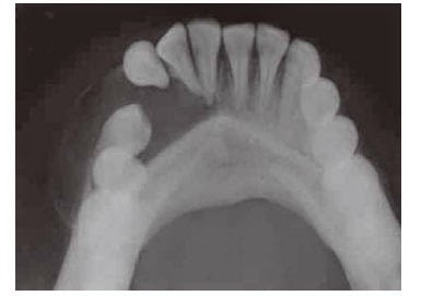
Radiological Features
AOT Radiologic feature
AOT of mandibular left canine region
image via: jaypeejournals.com
  • Radiographically adenomaroid odontogenic tumor presents a well-defined , unilocular,radiolucent area,which is often enclosing a tooth or tooth-like stucture.
  • Multiple small,radiopaque foci of varying radiodensity may be predent inside the lesion .
  • Expansion of the distrotion of the cortical plates and displacement of the roots of the adjoining teeth are sometime seen.
  • The border of the lesion is not well corticated and it consistently engulfs the impacted tooth including its root.This feature differentiates adenomataid adontogenic tumor from dentigerous cyts,since the later lession encloses only the crown portion of on impacted tooth.

Differential Diagnosis

  • Dentigerous cyts
  • Globulomaxillry cyts
  • Lareral periodontal cyts
  • Odontome
  • Unicystic ameloblastoma
  • Ossiffying or cementifying fibroma
  • Calcifying epithelial odontogenic tumor
  • Calcifyinh epithelial odontogenic cyts.


Histopathological Feature

  • Microscopically, adenomatoid odontogenic tumor reveals neoplastic odontogenic epithealial cells,proluferating in multiple "duck-like" patterns,within a thin but well-vascularized stroma.
  • The presence of these duck-like strucrures often give the lesion an adenomatoid or glandular apperance.
  • Each dick-like structure is bordered on the periphery by a single layer of tall columnar cell resembling ameloblasts.
  • Serial sectioning reveals that the lumens are blind ended ang they probably represent an abortive attempt at anamel oraga formation.
  • The lumen of the duck-like structures are filled with a homogenous eosinophilic coagulum.
  • Small foci of calcification are often seen , which are scattered throughout the lesion.
  • In some cases , the neoplastic cells are arranged in solid nests or rosettem and these cells simetime may fill up the entire lumen of few ducts.
  • Droplets of amorphous (PAS prositiv) eosino philic materials are frequently found in between the neoplastic calls.
  • In some adenomatoid odontogenic tumos tubular dentin or enamel matrix may also be found.
  • The neoplasm is almost always well-encap sulated and the connective tissue stroma may occasionally contain diffuse areas of hyaline materials.


Treatment 

By surgical enucleation . The associated tooth has to be removed and recurrence is rare.


Ameloblastoma Complete Info (with Pictures)

Definition
Ameloblaatoma is a benign locally aggressive neoplasm arising from the odontoenic epithelium and it is the most common odontogenic neoplasm of the oral cavity.

Etiology
Exactily not known however , the following factors may predispose the formation of ameloblastoma:

  • Trauma.
  • Infection.
  • Previous inflammation.
  • Extraction of tooth.
  • Dietary factors.
  • Viral infection.

Clinical Features

  • Incidence Approximately 1 percent among all oral tumous.
  • Age- second, third,fourth and fifth decade of life , the mean age of occurrence is about 32 years.This lesion occurs more commonly in blacks than whies.
  • Sex-  Males are affected more often females.
  • Site- Ameloblastoma in most of the cases involve the mandible (80%), especially in the molar-ramus area (70%) , although some lesions may develop in the premolar (20%) or symphysis (10%) regions.
  • Maxillary tumors also commomly involve its posterior part  and the lesions ofter have a tendency to invade into the antrum (15%) or the nasal floor.
  • Extraosseous ameloblastomas can rarely occur mostly in relation to the gingival.


Clinical Presentation
Ameloblastoma
 Ameloblastoma in right jaw
Via wikipedia.org

  • Clinically ameloblastoma commonly presents a slow enlarging,painless,ovoid or fusiform,bony hard swelling of the jaw.
  • The lesion causes expansion and didtortion of the cortical plates of the jawbone and displacement of the regional teeth; these are often leading to gross facial asymmetry.
  • Pain ,paresthesia and mobility of the regional teeth may be present in few cases.
  • Most of the be patients report with a typical long time history of presence of an “abscess” or a “cyst” in the jow bone that was operated on several occasions but has recurred after each attempt.
  • Lerger lesions of ameloblastoma often cause severe expansion ,destruction and thinning of the cortical plates , which often result in “fluctuation”or “egg shell cracling” of the affected bone.
  • Pathological fractures”,may occur in many such affected bones.
  • The mucosa overlying the tumor appears normal and the regional teeth are usually vital.
  • In some cases , smaller lesions may remain asymptomatic for a longer duration of time and are detected incidentally during routine radiographic examinations.
  • Many untreated lesions may reach to an enor –mous size with time.
  • Sometimes larger lesions may perforate the cortical plates and protrude outside the bone as a modular mass.
  • Maxillary tumors can invade into the maxillary air sinus and extend further up to the orbit or the nasopharynx;thereby or nasal obstruction,etc.
  • Some of the lesions may progress to ethmoidial air sinuses or even up to the cranial base.
  • Extraosseous ameloblastoma often produces a small ,nodular growth in the gingival.


Radiological Feature
Ameloblastoma
Right jaw ameloblastoma
Image via: Radiopedia.org


  • Radiographically ameloblastoma usually pre-sent a well-defined,multilocular ,radiolucent area in the bone with a typical “honey-comb” or “soap-bubble” appearance.Few lesion can be unilocular too.
  • The lerger lesions often couse expansion,dis-tortion or even perforation of the cortical plates.
  • In radiograph the lesion typically exhibits an irregular and “scalloped”margin.
  • Resorption of the adjoining normal teeth is often seen in rapidly growing lesions.
  • Ameloblastoma can cause expansion of the lower border of mandible. An irregular,”scallo-ped margin “ are often the typical features of ameloblastoma.
  • An the neoplasm progresses it sometimes become associated tooth (mostly the third molars) and in such cases the lesion may be resemble a dentigerous cyst.


Differential Diagnosis

  • Odontogenic keratocyst (Primordial cyst)
  • Dentigerous cyst
  • Central giant cell granuloma
  • Central hemangioma
  • Aneurysmal bone cyst
  • Fibromyxoma.


Macroscopic Features

  • On naked eye examination the tumor presents a cylindrical or fusiform swelling, which expands the bone so the severely that it can bebroken by digital pressure (egg-shell crrackling).
  • Perforation of the bone with subdequent protrusion of the tumor outside the bony wall is often noticed.
  • Cut section of ameloblastoma often appears as a “grayish-white”mass ,which contains some ‘cyst-like ‘ spaces. However no calcified is usually found within the tumor.
  • Some lesions are made up entirely of solid tissue mass although most of them have some cystic spaces of varying size within them.
  • Some intratumor cysts are large and contain either a straw colored fluid or a semi solid gelatinous material.
  • Sometimes one or two teeth may be present within thw lesion.


Histopathological Features
Ameloblastoma Microscopic
Microscopic view :Ameloblastoma
Image via: Wikipedia.org

Histologically ameloblastoma shows neoplastic proliferation of odontogenic epithelial cells mostly in two distinct patterns ; (i) Plexiform type and (ii) Follicular type.

Plexiform Ameloblastoma

  • In this variant of ameloblastoma the neoplastic odontogenic epithelial cella proliferate in the form of “ contimuous anastomosing strands”.
  •  This patterm of neoplastic cell proliferation is also often called a “ fishnet like” pattern of arrangement.
  • The peripheral layer of cells tall columnar in nature and they often resemble the amelo-blasts.
  • Reverse polarization of the nucleu of these bordering cell is indistinct.
  • The cells situated at the center portion of the strands often resemble the stellate reticulum cells;while the cells located  berween the columnar cells and stellate reticulum cells often resemble the startum intermendium.
  • The intervening connective tissue stroma is usually thin , with minimum cellularity and often it show multiple areas of cystifications , which may be either large or small in size .


Follicular Ameloblastoma 


  • In follicular type , the neoplastic odontogenic epithelial cells proliferate in the from of multiple , discrete , follicles or islands within the fibrous connective tissue stroma.
  • Each follicle-like structure is bordered on the periphery by a single layer of tall columnar cells resembling ameloblasts.these cells have well-defined neclei situated away from the basement are called “reverse polarization” of the cell nuclei.
  • The cells located at the center of the follicles are loosely arranged and are triangular in shaps;these cells are widely seiarated from one another and they often resemble stellate reticulum cell (normally seen in the bell stage of odontogenesis)
  • While the cells located in berween the peripheral and the central group of cells appear as the stratum intermedium.
  • Occasionally a distinctive zone of hyalinization is seen surrounding the follicles.
  • Microcyst formation is often observed inside these follicles and the cysts sometimes may be large enough to occupy the entire inner part of the follicles.
  • Most of the follicular ameoloblastomas exhibit cyst formation particularty if the lesion is large.
  • The intervening connective  tissue strimas is delicate in nature and it consists chifly of collagen bundles,fibroblasts and blood vessels etc.
  • Extraosseous ameloblastoma consists of basaloid cells or they may even resemble the conventional intraosseous ameloblastomas.


Other Histological Types of Ameloblastoma

Besides the plexiform and the follicular types some other histological types of ameloblastomas can occur and they are as follows :

Acanthomatous type of ameloblastoma: It occurs in relation to follicular ameloblastoma and in this type the stellat reticulum-luke cells at the center of the follicles undergo squamous metaplasia. Sometimes the neoplastic epithelial cells can even produce “keratin pearls” whith the follicle the meoplastic cells may exhibit individual cell keratinization.

Granular cell type of ameloblastoma: In this type the cytoplasm of the stellate reticulum like cells and even the ameloblast like cells appear swollen and the cells are often densely packed with multiple, coarse,eosinophilic granules.Histologically this lesion often resembles “granular cell myoblastoma” and ultrastructural studies indicate that these geanules are either lysosomal elements or residual bodies.

Besal cell type of ameloblastoma: this lesion shows excessive proliferation of cuboidal shaped,bassaloid cells in narrow strands with the absence of stellate reticulum or other located cells The tumor often resembles basal cell carcinoma.

Cystic type of ameloblatoma: these lesions often exhibit multiple,small,microcyst formation inside the tumor.Some of the cysts are large in size

Desmoplastic type: In this type the epithelial islands or the strands are amall in size and the cells are ceboidal in shape and darkly stained.The cells of the epithelial components are widely separated by dense fibrous tissue. In this cells often penetrate into the surrounding trabacular bene.

Histogenesis of Ameloblastoma

Ameloblastoma develops from the odontogenic epithelial cells or their remnants but the exact cell of its origin is not very clearly known.According to different investigators, the possible cells or tissues from where ameloblastoma may arise are as follows.

  • Enamel organ of the developing tooth gern 
  • Cell rest of Serre (remnsnts of dental lamina)
  • Epithelial lining of the odontogenic cysts especially the dentigerous cyst.
  • The basal cell layer of the oral epithelium (rarely)
  • Cell rest of malassez.

Treatment

Surgical enucleation of the tumor and thorough curettage of the surrounding bone.Sometimes radical surgical approach may have to be adopted in case of repeared recurrences of the lesion Some tumors may cause distant matastasis.


Odontogenic Tumor : Defination & Classification

Definition: Odontogenic Tumors or neoplasms are a complex group of lesions derived from the dental formative tissues or their remnants ( tissues associated with the development of tooth and its supporting structures ). The constituent tissues in each of these neoplasms can resemble the various tissues found during normal odantogenesis,from inception of the tooth germ to tooth eruption.

The tooth formation or odontogenesis begins in the 6th week intra-uterin life and it originates from the oral epithelium covering the maxillary and mandibular alveolar processes .During the initial period “bud-like” swellings appear from the basal layer of the oral epithelium at specific location where individual teeth will appear from in future .


CLASSIFICATION OF ODONTOGENIC NEOPLASMS (TUMORS)

Benign Odontogenic Neoplasms 

Ameloblastoma
Ameloblastoma a kind of odontogenic tumor in left jaw:
Case courtesy of Dr Frank Gaillard, Radiopaedia.org
1.  Neoplasms of epithelial tissue origin 
a. Ameloblastoma
b. Squamous odontogenic tumor
c. Calciflying epithelial odontogenic tumor (CEOT)
d. Clear cell odontogenic tumor.
2.  Neoplasms of mixed tissue origin (Made up of both epithelium and mesenchymal tissues)
a. Adenomatoid odontogenic tumor (AOT)
b. Ameloblastic fibroma
c. Ameloblastic fibro-odontoma / fibroden-tinoma
d. Odonto-ameloblastoma
e. Complex odontoma
f. Compound odontoma
g. Calcifying epithelial odontogenic cyst.
3.  Neoplasms of the mesenchymal tissue origin 
a. Odontogenic fibroma
b. Odontogenic myxoma
c. Cementoma
d. Famillial gigantiform cementoma
e. Cementifyin fibroma
f. Bening cementolastoma

Malignant Tumors

1.  Odontogenic carcinomas :
a. Malignant ameloblastoma
b. Primary intra-alveolar carcinoma
c. Malignant variants of other epithelial tissue neoplasms
d. Malignant changes in odontogenic cysts.
2.  Odontogenic sarcomas :
a. Ameloblastic fibrosarcoma
b. Ameloblastic carcinosarcoma
c. Ameloblastic fibrodentinosarcoma

Neoplasms of Debatable Origin 
Melanotic neuro-ectodermal tumor of infancy 
Congenital gingival granular cell tumor (congenital epulis)

Can Xylitol Chewing Gum Protect Your Teeth?

Xylitol
image via: Vitalsurge.com
You have always heard that eating sugar candy is bad for your teeth. but what if i say "chew some gum and protect your teeth"

Yes you are hearing right, i am suggesting you chewing gum ..but how does it help protect your teeth?..

I am suggesting you to have SUGAR FREE Xylitol containing chewing gums present in the market in various brands.


How does it help.?
As I have discussed earlier about the process of Caries/Decay, the organism responsible for decay of the tooth is S mutans.It promotes an acidic environment in our mouths which leaves enamel susceptible to damage, erosion and eventually cavities.

Luckily, nature has provided a cheap, natural and safe remedy for this problem: xylitol. xylitol is a sweet substance commonly found in birch trees and in the fibrous portions of many fruits and vegetables.I want to discuss its application as a cavity fighting agent.

A recent study out of Korea examined the effect on cavities caused by the regular chewing of xylitol sweetened gum.

Two groups of women were assigned to chew either regular or xylitol gum for a period of one year. At ten points during that year, saliva samples were taken from these women and analyzed to determine the amounts of S. mutans.

In the xylitol gum group, the levels of S. mutans decreased consistently as the study progressed.  The researchers also found that the S. mutans produced a lower amount of sticky substances in the xylitol chewers. This is relevant because the stickiness allows for acids to cause more damage to our teeth. The combined effects of  chewing the xylitol gum led to an oral environment that was less prone to cavity formation.

The results of this first study were encouraging. But I wanted to see if this might be a fluke. It seems that it is no

Facts & Figures-
In December of 2008, a review appeared in the Journal of the American Dental Association. In it, the researchers examined the findings of 19 studies relating to the use of xylitol and sorbitol gums in the prevention of tooth decay. Their analysis found that the xylitol gum studies showed the greatest cavity prevention.

Here’s a breakdown of a few different sugar alcohols and their overall preventive effect:
Xylitol Gum – 58.66%
Xylitol & Sorbitol Combination Gum – 52.82%
Sorbitol Gum– 20.01%
Sorbitol & Mannitol Combination Gum – 10.71%

As you can see, the xylitol component appears to be the most important factor in the promotion of oral health.

So how exactly does xylitol help protect teeth? It is believed that it works to starve harmful bacteria, like S. mutans. This leads to a less acidic environment that is less prone to decay and plaque formation.

Calcium added Xylitol gums-
Preventing cavities is a very positive thing. But is there a way to strengthen enamel, if it’s weak to begin with? Maybe so.
A few years ago, scientists in Japan published a study that tested a combination gum that included calcium lactate and xylitol. Their aim was to see if such a gum could actually make tooth enamel stronger.

Volunteers were asked to either a) chew no gum, b) chew xylitol gum or c) chew gum with xylitol and calcium lactate. The voluteers chewed 4 pieces of each gum for 2 weeks. After which, their enamel was measured using an X-ray.

The results showed that the xylitol-calcium gum was about 50% more effective in promoting remineralization than the xylitol-only gum. The authors concluded that, “chewing gum containing xylitol + calcium lactate could enhance remineralization of enamel surface”.
So i suggest having these xylitol chewing gums instead of sugar candies. Care for your teeth they are precious.


Top 10 qualities to be a successful Dentist

Dentist
Mediaphotos : Getty images
In..today's changing world where most of the people are taking career in dentistry as a nice paying profession some light has to be focused on the qualities we must posses qualities to be a successful Dentist.

I want to call this 'unfortunate' as we don't have any training sessions to develop these qualities in our dental syllabus, these qualities must be cultivated by own.

At this high time when dental colleges have  mushroomed every where and thousands of dentists coming out every year and there is 'so called raising competition'  if you really want to make difference then having a degree is not enough.

I call it "Passion" you have to be passionate about dentistry.Ask yourself Will I do it even if I don't get paid? Do I like it so much? if answer is yes then 'You are Passionate enough' .

Here are some qualities you need to develop. 

1. Has a Good Manner: A great medical professional has a good manner and makes patients feel comfortable and at ease during exams and treatments.

2. Has a Sense of Empathy: A great medical professional has a strong sense of empathy and understands what it is to feel pain and suffering. They are supportive and have a genuine interest in improving a patient's well-being.

3. Has Great Communication Skills: A great medical professional has excellent communication skills. They can explain complicated medical terminology in laymen's terms to the average patient. They also have excellent listening skills and take the time to understand what a patient's needs are.

4. Has Sharp Problem Solving Skills: A great medical professional has excellent problem solving skills and can quickly determine solutions to problems. Working in health care, by definition, involves solving problems of the human body.

5. Is Always Very Thorough: A great medical professional is always very thorough in their work. They recognize that the smallest oversight can have grave consequences and therefore are sure to cover all the bases in everything they do.

6. Offers Support for Patient Decisions: A great medical professional acts as a partner with a patient in treatment decisions and understands that ultimately, all decisions lie in the hands of the patient. They offer full support of patient decisions.

7. Offers Time to Patients: A great medical professional spends adequate time with their patients and never rushes through an exam during a busy day. They give each patient enough time to make a proper diagnosis or to offer a thorough treatment.

8. Possesses Significant Knowledge: A great medical professional has extensive knowledge of the human body and its ailments. They are not afraid to admit when they do not know something and will either research it or refer a patient to someone better qualified.

9. Possesses Strong Sense of Ethics: A great medical professional has a strong sense of ethics and never compromises their integrity or values.

10. Pursues Continuing Education: A great medical professional recognizes that the medical field is full of new research and developments, and they stay on top of everything new in the field. They read research journals and take training classes to stay current.

What is Sialolithiasis –Definition, Pathogenesis, Clinical features, Treatment, Histopathology, Sialography.

Sialolithiasis

Definition- sialolithiasis is a pathological condition, characterized by the presence of one or more calcified stones (sialliths) within the salivary gland itself or within its duct.

Pthogenesis

The exact mechanism of formation of sialolith is not known. It is generally believed that initially a small and soft nidus forms within the salivary glands or its ducts due to some known reasons.

The nidus is made up of mucin, protiene acteria and desquamated epithelial cells.

Once a small nidus frms, it allows concentric lamellar crystallizations to occur due to the precipitation of calcium salts.

The sialolith increases in size with time as layer after layer o salts become deposited , just like growth rings in a tree.

Small sialoliths can be expelled in the mouth with the salivary secretions but bigger sialoliths continue to expand until a duct is completely closed.

It is important to note that the formation of sialolith is more common in relation to the submandibular gland and it’s duct.

sialolithiasis

Clinical features

Age- Sialolithiasis usually occurs among the middle aged adults, however, some cases are reported in children.
Sex- There is slight male predominance.
Sites- 70% in submandibular salivary gland and its ducts. Parotid gland is next

Clinical Presentation-

  • In many cases sialoliths do not produce any symptoms and are detected only on routine radiographic examination.
  • Chief complain ; intermittent pain, recurrent sumbandibular swelling, discomfort during meals.
  • Pain is drawing or stinging in mild cases but stabing and sharp in severe cases.
  • Stone in the duct can be palpated by bimanual palpation with both the fingers.
  • During examination the flow and clearness of the saliva should be checked.
  • Persistent swelling of the duct due to chronic obstruction by the sialolith eventually leads to chronic sclerosing sialadenitis.
  • Multiple stone or bilateral cases of sialolithiasis can also be seen.
  • Formation of fistulas, sinus tract, ulcerations in the area may develop in chronic cases.
  • Necrosis of gland acini and lobular fibrosis may occur which results in complete loss of secretion from the gland.
  • Parotid stones often cause firm swelling over the ramus of the mandible, the swelling over the ramus of the mandible, the swelling also increases during meals.

How Sialolithiasis is diagnosed
  • Radilography of sialolithiasis
  • Submandibular sialolithiasis are easily detected by mandibular standard occlusal radiograohs which typically disclose the presence of calcification in the floor of the mouth.
  • Whn a sialolith is located in submandibular gland area then a lateral jaw radiograph will be helpful in location the exact position of the stone.
  • Panaromic radiographs usually detect the parotid gland stones.
  • If only branches of the submandibular gland duct are affected a posterior occlusal film submento vertex and some time a lateral jaw film may be required.

 Sialography of sialolithiasis
  • Sialogrohy refers to the method by which detection of salivary stones within the glad or its duct is done by giving a retrograde injection of a radiopaque dye within the  duct system and obtaining a radiograph thereafter in order to see the size and distribution of the sialolith.
  • Ultrasonograhy and CTscan
  • These are also excellent non invasive technique useful in detecting the sialoliths

Macroscopic appearance
  • On gross examination, sialoliths appear as round or oval rough or smooth solid masses, which vary considerably in their size.
  • These stones are heavily calcified and are often multi nodular although some stones are found in small aggregates.
  • The color of the stone is usually yellowish or yellowish white.


Why Are My Teeth Getting Stained ?

Question- I go to my dentist regularly and get my oral prophylaxis & cleaning done in every 6 months, My teeth become pearly white after cleaning and their are no stains but they start getting stained in no time ! I can see visible stains after 3 weeks.Why are my teeth getting stained even when I brush properly.

Although teeth staining have multiple causes but the above case is a simple case of poor oral hygiene practice. I'm not saying that the person is not will to keep his teeth clean but he is actually practicing wrong oral hygiene techniques like not knowing proper brushing technique, not flossing and brushing only once in the morning.

Cause of teeth getting stained after scaling and cleaning:
teeth getting stained
Stained between teeth Img: www.smileartistry.com.au

After professional cleaning surface of your teeth become absolutely clean but as you start eating a soft Dental pellicle start to form on your teeth.It is a protein film that forms on the surface enamel by selective binding of glycoproteins from saliva.

When not cleaned and flossed properly oral bacteria start to make colonies on this pellicle layer and hence they for a layer called as Plaque. This plaque can take the colors of foods and beverages your consume and it looks like a staining on your tooth.when you get your teeth scaled and cleaned at the dentist he removes this plaque. When people does not get this Plaque cleaned and it keep on accumulating it becomes Calculus which is a calcified structure.

Foods & Habits That Cause Your Teeth to Get Stain


Tea Or Coffee:
Tea Or coffee contains such kind of plant extracts that have very high tendency of sticking as stains on your teeth. No matter what type pf tooth paste you use, no matter what type of brushing you do, if you are consuming tea or coffee more then once a day every day in a year then your teeth will catch stain.Either get a professional cleaning or change your habit.

Smoking: Tobacco smoke contains nicotine and tar these two ingredients are the main culprit for the smoking stains. tar has a black staining and nicotine has a yellowish staining property. So a smoker will have a blackish and line at the junction of the gums and tooth and yellowish hue on the teeth. Leave the habit, Get your teeth cleaned.

Red Wine- Red wine is made up of grapes which have natural colors these natural colors will bind with the tooth surface and result in teeth staining, but this process is low and mild and occurs to them who consume red wine regularly.

Some Cold drinks and Energy drinks: Cold drinks and energy drinks have coloring agents in them. These agents can deposit on the plaque of the tooth and then get solidify.

Some berries and veggies: I really don't think that veggies should be blamed for the staining of teeth but clinically it have been seen that there are some berries and veggies which have colored juices in them which can stick to the plaque and impart

Solution to Get Rid of Repeated Teeth Staining

Avoid stain producing habits and food.
Brush twice a day with correct brushing technique.
Use floss once a day.
Use inter dental brush to clean gaps between teeth.
Get oral prophylaxis done every 6 months.
Keep oral hygiene good.